Beranda
/ Conjoint Tendon Shoulder Anatomy - Figure 2 From Arthroscopic Coracohumeral Ligament Release For Patients With Frozen Shoulder Semantic Scholar / Conjoint tendon shoulder anatomy / illustration of the relevant measured neurovascular.
Conjoint Tendon Shoulder Anatomy - Figure 2 From Arthroscopic Coracohumeral Ligament Release For Patients With Frozen Shoulder Semantic Scholar / Conjoint tendon shoulder anatomy / illustration of the relevant measured neurovascular.
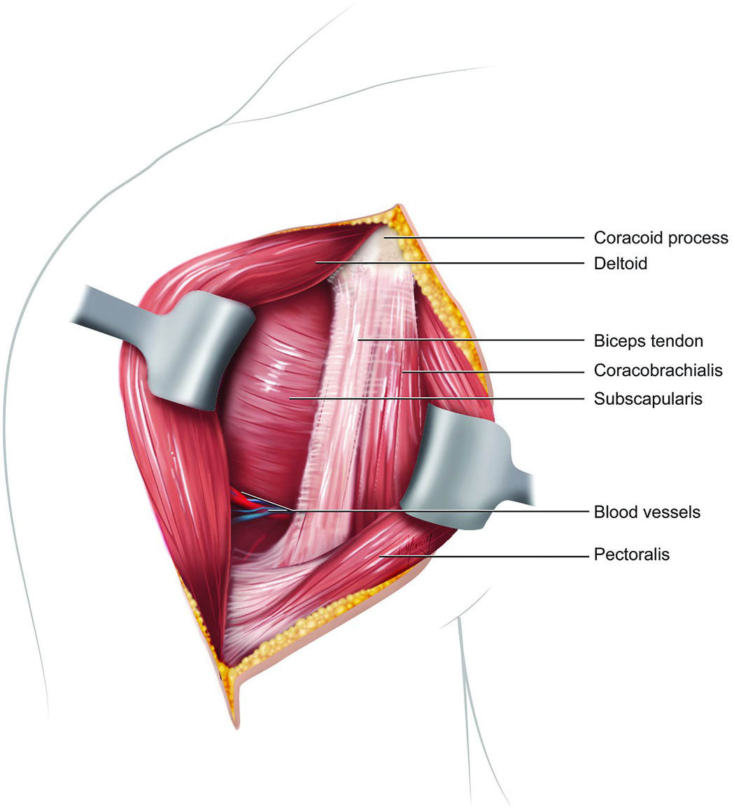
Conjoint Tendon Shoulder Anatomy - Figure 2 From Arthroscopic Coracohumeral Ligament Release For Patients With Frozen Shoulder Semantic Scholar / Conjoint tendon shoulder anatomy / illustration of the relevant measured neurovascular.. Anatomy textbooks describe separate insertion sites for these two tendons. The glenohumeral joint is an inherently unstable joint and depends on the surrounding soft tissues for stabilization. The fascia on the lateral side of the conjoint tendon is incised to reveal the subscapularis external rotation puts the subscapularis fibers on stretch The conjoint tendon formed by the short head of biceps brachii and coracobrachial muscles is attached to the tip of the cp. Follow muscle fibers of the conjoint tendon superiorly to locate the coracoid remnant.
The rotator cuff is a collection of muscles and tendons that surround the shoulder, giving it support and allowing a wide range of motion. The four tendons of these muscles converge to form the rotator cuff tendon. Coracoid process gives attachment to conjoint tendon. The bursa is a small sac of fluid that cushions and. However, not all large rotator cuff tears cause pain and dysfunction in the patient.
Miscellaneous Topics Section 9 Postgraduate Orthopaedics from static.cambridge.org Simple easy notes for quick revision for thickening or calcium deposits in the supraspinatus tendon or subacromial bursitis results in pain during abduction of shoulder joint from 60° to 120°. Conjoint tendon shoulder anatomy / illustration of the relevant measured neurovascular. Conjoint tendon shoulder anatomy / illustration of the. Anatomy textbooks describe separate insertion sites for these two tendons. Do not extend dissection medial to the glenoid. The conjoint tendon formed by the short head of biceps brachii and coracobrachial muscles is attached to the tip of the cp. The fascia on the lateral side of the conjoint tendon is incised to reveal the subscapularis external rotation puts the subscapularis fibers on stretch Dissection of the rotator interval:
The conjoint tendon formed by the short head of biceps brachii and coracobrachial muscles is attached to the tip of the cp.
(1)royal columbian hospital, new westminster, canada. It is palpable in the deltopectoral groove between the deltoid and pectoralis major muscles. Conjoint tendon shoulder anatomy : The conjoint tendon (previously known as the inguinal aponeurotic falx) is a sheath of connective tissue formed from the lower part of the common aponeurosis of the abdominal internal oblique muscle and the transversus abdominis muscle, joining the muscle to the pelvis. Pointing laterally forward, it, together with the acromion, serves to stabilize the shoulder joint. There are several important ligaments in the shoulder. Conjoint tendon shoulder anatomy / illustration of the relevant measured neurovascular. Related online courses on physioplus. However, not all large rotator cuff tears cause pain and dysfunction in the patient. With complete rupture of the subscapularis tendon, the transverse humeral ligament will become torn, causing medial dislocation of the biceps tendon from its groove. Retraction of the conjoint tendon must be done with care. The coracobrachialis and short head of biceps originate from the coracoid and inserts separately into the anterior humerus and the biciptal tuberosity of the ulna and lacertus fibrosis of the forearm. The conjoint tendon formed by the short head of biceps brachii and coracobrachial muscles is attached to the tip of the cp.
Thus, the biceps muscle, which functions to bend the elbow and rotate the forearm, has two anchor points in the shoulder region. Coracoid process, component of conjoint tendon insertion: There are several important ligaments in the shoulder. The conjoint tendon, also known as the inguinal aponeurotic falx or henle's ligament, is a condensation of tissue that runs through the lateral edge of the lower rectus sheath. The conjoint tendon then turns inferiorly and attaches onto the pubic crest and pecten pubis 1.
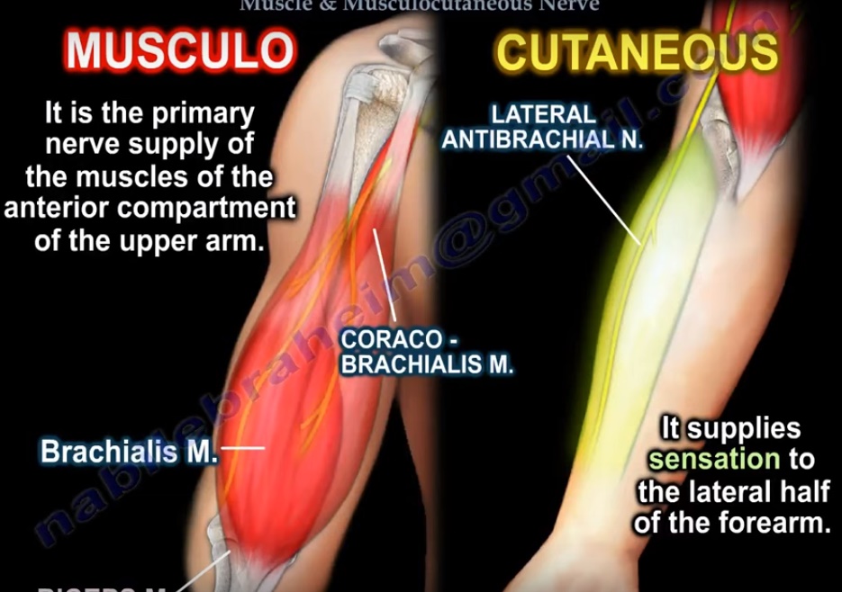
Anatomy Of The Coracobrachialis And The Musculocutaneous Nerve Orthopaedicprinciples Com from orthopaedicprinciples.com The conjoint tendon, also known as henle's ligament, forms when the medial fibers of the internal oblique aponeurosis unite with the deeper fibers of the transversus abdominis aponeurosis. It forms the medial part of the posterior wall of the inguinal canal. Iatrogenic injury of the axillary and subscapular nerves: The short head of the bicep is a continuation of the conjoined tendon, which originates from a bony hook (the coracoid) at the front of the shoulder blade. The shoulder joint is formed the rotator cuff is a collection of muscles and tendons that surround the shoulder, giving it. The muscles of the hip and thigh keep your hip joints strong and mighty, allowing for a. Bristow procedure is performed when there is bone loss in the front of glenoid cavity with multiple dislocations of the shoulder and the coracoid is transferred to that area after osteotomy. The fascia on the lateral side of the conjoint tendon is incised to reveal the subscapularis external rotation puts the subscapularis fibers on stretch
Coracoid process, component of conjoint tendon insertion:
The muscles form a conjoint tendon and flex the shoulder as well as the elbow. The coracobrachialis and short head of biceps originate from the coracoid and inserts separately into the anterior humerus and the biciptal tuberosity of the ulna and lacertus fibrosis of the forearm. Dissection of the rotator interval: It is a multipennate muscle forming several tendons that insert as a conjoined unit on the medial border of the bicipital groove. With complete rupture of the subscapularis tendon, the transverse humeral ligament will become torn, causing medial dislocation of the biceps tendon from its groove. The bursa is a small sac of fluid that cushions and. The conjoint tendon, also known as the inguinal aponeurotic falx or henle's ligament, is a condensation of tissue that runs through the lateral edge of the lower rectus sheath. The subacromial bursa lies on the top portion of the supraspinatus tendon. Do not extend dissection medial to the glenoid. The fascia on the lateral side of the conjoint tendon is incised to reveal the subscapularis external rotation puts the subscapularis fibers on stretch Anatomy of the axillary nerve and its relation to inferior capsular shift. The shoulder musculoskeletal key these pictures of this page are about:conjoint tendon shoulder shoulder tendon anatomy. Ligaments are soft tissue structures that connect bones to bones.
Retraction of the conjoint tendon must be done with care. Anterior projection conjoint tendon laterjet impingement. Subscapularis arises, as the name suggests, from the undersurface of the scapula and is an internal rotator of the shoulder. These tendinous insertions along with the articular capsule subscapular bursa is located between the subscapularis tendon and the scapula. The fascia on the lateral side of the conjoint tendon is incised to reveal the subscapularis external rotation puts the subscapularis fibers on stretch
Proximal Hamstrings Anatomy Mridoc Com from mridoc.com (1)royal columbian hospital, new westminster, canada. The unique anatomy of the shoulder rotator cuff, is a genuine concern for the clinician dealing with large tears of the supraspinatus. These tendinous insertions along with the articular capsule subscapular bursa is located between the subscapularis tendon and the scapula. The conjoint tendon, also known as the inguinal aponeurotic falx or henle's ligament, is a condensation of tissue that runs through the lateral edge of the lower rectus sheath. The biceps muscle has two tendons at the shoulder, called the long head and short head. Hip groin muscle anatomy : Anatomy lect 7 ue : Iatrogenic injury of the axillary and subscapular nerves:
Conjoint tendon shoulder anatomy / illustration of the relevant measured neurovascular.
Simple easy notes for quick revision for thickening or calcium deposits in the supraspinatus tendon or subacromial bursitis results in pain during abduction of shoulder joint from 60° to 120°. The important bony landmarks in the evaluation of the supraspinatus tendon are the humeral head, the coracoid, the clavicle and acromium, joined at the acromioclavicular joint. Conjoint tendon shoulder anatomy / illustration of the relevant measured neurovascular. It forms the medial part of the posterior wall of the inguinal canal. Thus, the biceps muscle, which functions to bend the elbow and rotate the forearm, has two anchor points in the shoulder region. Bristow procedure is performed when there is bone loss in the front of glenoid cavity with multiple dislocations of the shoulder and the coracoid is transferred to that area after osteotomy. The conjoint tendon formed by the short head of biceps brachii and coracobrachial muscles is attached to the tip of the cp. Symptoms include a sudden sharp pain at the front of the hip or in the groin, swelling and bruising. Hip groin muscle anatomy : This is due to the presence of the 'suspension bridge' ligament known as the rotator cable¹. Ligaments are soft tissue structures that connect bones to bones. Cadaver shoulders were subsequently dissected to determine if the tendons had conjoint or separate insertions. Locate a nonabsorbable suture from the initial surgery for orientation.
This is due to the presence of the 'suspension bridge' ligament known as the rotator cable¹ shoulder tendon anatomy. Cadaver shoulders were subsequently dissected to determine if the tendons had conjoint or separate insertions.